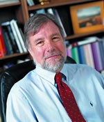
Michael Beattie, PhD
Background: The CNS response to injury shares features with other organ systems, and includes surprisingly robust repair mechanisms. The cascade of events that leads to dysfunction and partial recovery after spinal cord injury, for example, includes overlapping degenerative and regenerative processes mediated by inflammation, excitotoxicity, growth and trophic factor production in all of the cellular components of the CNS including neurons, glia, immune cells, and vascular elements. In this complex cascade, neural connections may be broken and remade and the final functional outcome reflects the resolution of all of these interacting events. We think that a better understanding of the biology of the lesion and repair processes will lead to better strategies for providing protection against secondary injury, enhancing self-repair, and for engineering repair through the use of cellular implants and biomaterials. We also are invested in the production of useful in vitro and in vivo testing systems for bringing these strategies into the clinic. The laboratory is co-directed by Dr. Jacqueline Bresnahan (Neurosurgery), and has recently moved from The Ohio State University to UCSF (November, 2006).
Major Goals: Our goal is to better understand the biological processes involved in CNS damage and repair so that we can help to develop effective strategies for treatment of CNS disorders, including spinal cord injury.
On-going research:
Preclinical animal models of CNS injury. Our group is known for developing preclinical models to study recovery of function after spinal cord injury, and for studies of the biology of neural injury and repair. Current projects include analyses of the interrelationship of post-injury inflammatory events and excitotoxic cell death, the roles of oligodendrocyte death and replacement in recovery after injury, and the development of stem and progenitor cell transplantation strategies for promoting recovery after spinal cord injury. The laboratory continues to develop new animal paradigms for modeling human neurotrauma. BASIC’s location at the SFGH along with the physician-scientists of UCSF Neurotrauma program will promote basic and clinical science interactions, and new animal models will be generated based on knowledge of current treatments and outcomes in the human neurotrauma arena. For example, there is an emerging interest in post injury edema, studied in rodents using high field MR imaging. Acute studies of interventions that can reduce edema in animal studies may be taken directly to clinical studies in the neurotrauma ICU. Our goal is to help translate the laboratory’s expertise in the biology of injury and recovery to treatments that can be implemented and tested in neurotrauma patients at SFGH and other centers.
Spinal cord degeneration, regeneration, and cellular therapies:
Transplantation of stem-like and precursor cells into spinal cord contusion lesions: This is currently a major thrust in the laboratory. Cells derived from a transgenic rat with ubiquitous expression of the human placental alkaline phosphatase (hPlAP) gene are isolated form embryonic spinal cord and treated with growth factors to produce a glial restricted precursor population. Transplantation into our well-established models of spinal cord injury allows assessment of survival, phenotype, and possible effects on a variety of neurological outcome measures. These cells survive even when transplanted immediately after injury, and some form astrocytes and oligodendrocytes. The rodent models in our laboratory are also suitable for proof of concept studies using human ES cells (see future directions). Collaborators include Mark Noble, Margot Mayer-Proschel, and Chris Proschel (University of Rochester).
Demyelination and remyelination:
We continue to study secondary degeneration of axons and the effects on oligodendrocytes and myelin. The hypothesis is that long-term apoptotic death of myelinating cells reduces the efficiency of conduction in spared axonal tracts after injury, and that saving these cells and/or producing remyelination through stimulation of endogenous stem cells or implantation of glial precursors will improve recovery from CNS injury. The secondary cell death may be due to a variety of receptor-mediated cascades, e.g. the activation of TNF receptors and p75 by endogenous ligands (e.g. Beattie et al, 2002). Recently, we have discovered that axonal degeneration can induce oligodendrocyte precursor cell (OPC) proliferation and differentiation. However, in the presence of oxidative stress, new cells are apparently lost via apoptosis. The role of microglial activation in these events is being studied both in vitro and in vivo. These projects overlap with those on inflammation and excitotoxicity, and also have relevance for other neurological disorders, including multiple sclerosis and stroke.
Inflammation and excitotoxicity:
We have found that the pro-inflammatory cytokine, TNF- a , enhances glutamate-receptor-mediated excitotoxic cell death in neurons in vitro and in vivo. This is due, at least in part, to changes in AMPA receptor trafficking to the cell surface (see E. Beattie et al, 2002). Confocal microscopy and biochemical techniques have been developed to track surface expression of these receptors in vivo after injury or nano-injections of injury-related substances.
